Chapter 1: The science and the scan
"Boring, But Necessary"
- Hybrid Imaging System
- Anatomic & Functional Exam
- The Machine
- “Whole Body” vs. “Skull Base to Mid-Thigh”
- Three Sets of Images
Produced
- Glucose Analog
- Malignancy & Glucose Metabolism
- Mechanisms for Increased Intracellular Glucose
- Why 18F-FDG Works
- Whole Body Assessment
- The “You’re Kidding Me!” Effect
- Post-Therapeutic Scar vs. Active Malignancy
- Detecting Malignancy
- Staging Malignancy
- Assess Response to Therapy
- Detecting Recurrence
- Not All Cancer is FDG-Avid
- Normal FDG-Uptake vs. Pathologic Uptake
- Technical Limitations
- Poor Patient Preparation
- Misregistration
- Brown Fat Activation
- SUV Problems
- Fields of View Discrepancy
- PET/CT Artifacts
- Timing of Exam After Therapy
- CT Images
- Non-Attenuation Corrected Images
- Attenuation Corrected Images
- Maximum Intensity Projection (MIP)
- Fusion of Images
- All Images Viewed in 3 Planes
7. Contrast Media: Oral & I.V.
- Who Gets Oral Contrast?
- Who Gets IV Contrast?
- Oral Contrast “Cocktail” Recipe
8. What the Patient Should Expect
- Documenting Height & Weight
- Private Resting Room
- Drinking PO Contrast
- FDG Injection
- Delay Between Injection & Scan
9. Safety Concerns with PET/CT Imaging
- Radiation Exposure to Patient
- Radiation Exposure to Patient’s Contacts
- Patient Contact with Pregnant Women
- Breastfeeding
Chapter 2: PET/CT Problems Which Limit Interpretation
"Something just doesn't seem right here"
- Optimizing Glucose & Insulin Levels
- Fasting Prior to Exam
- Diabetic Patients
- Low Carbohydrate Diet
- Hydration
- Strenuous Exercise
- Voiding Prior to Exam
- Patient Instruction Sheet
- Definition
- Distribution / Appearance
- Don’t Miss the Hidden Nod
- Reporting Language
- Prevention
3. Timing of PET/CT Exam After Therapy
- “Rule of 3”
- Chemotherapy: 1 month
- Surgery: 2 months
- Radiation: 3 months
- Etiology: Hybrid Imaging
- Patient Movement
- Respiratory Motion
- Breathing Techniques
- Bowel Peristalsis
- False Positives
- False Negatives
- Reporting Language
- Beam Hardening
- Diaphragmatic Mismatch
- Linear Hand Motion
- Attenuation Correction
- Differing Fields of View
- Poor Patient Preparation
- FDG Extravasation
- Extensive Brown Fat
- Metformin-Induced Bowel Uptake
- Marked Reactive Marrow Uptake
- Extensive Tumor Uptake in
- Different Types of SUV Measurements
- Factors that Influence SUV Measurements
- What SUV Number Indicates Malignancy?
- What Percent Change in SUV on a Follow Up Exam is Significant?
- How to Compare Exams With Very Different Background Metabolic Activities?
Chapter 3: The Standardized Uptake Value (SUV)
"The good, the bad & the ugly"
1. What is the SUV & Why Used?
- Quantitative vs. Qualitative Assessment
- Unitless Measurement
- Formula
- SUV = ?
- Patient Preparation
- Time Between FDG Injection & Scan
- Partial Volume Effects
- Extravasation
- Patient Weight
- Size & Position of ROI
- Attenuation Correction Artifacts
- Consensus?
- Body Weight
- Lean Body Mass
- Ideal Body Weight
- Body Surface Area
- Maximum vs. Mean
- Average SUV’s by Organ
4. Interpreting the SUV: Threshold Values, “Oncologic Plausibility” & Relative Uptake
- Precise Threshold Values?
- “Oncologic Plausibility”
- Relative Uptake
- Assessing Nodes in Lymphoma Cases
- Assessing Nodes in Non-Lymphoma Cases
- Potential Lesions in Solid Organs
- Pulmonary Nodules
5. What % Change in SUV on a Follow Up Exam is Clinically Significant?
- The Problem
- Current Recommendations
6. How to Compare Sequential Exams With Very Different Background Activities?
- Differing Background Metabolic Activities
- When Qualitative Assessment is Required
- Reporting Language
7. Should We Just Abandon the SUV?
- Pros & Cons
- “Qualitative” Definitions
- Mild
- Moderate
- Intense
- Final Recommendations
Chapter 4: Our Systematic Approach to Reading a PET/CT
"Eat your vegetables"
1. Reading Station & Reading Software
- Reading Station
- Monitor Set-Up
- PET/CT Reading Software
- Hanging Protocol
- Reading in Context (“Oncologic Plausibility”)
- Measure Size on CT, Not on PET Images
- Abnormality Seen Only on First PET Image
- Assess the Patient’s Main Pathology Last
- Beware the Ureter
3. Excellent Views: The MIP, Coronal & Sagittal Images
- 3-D Rotating MIP & Coronal “Quick MIP”
- Coronal PET
- Sagittal PET
4. Written Annotations While Reading
- Numbers, Numbers & More Numbers
- Size & SUV Annotation System
- Sample Annotation Sheet
- Goals of Reporting
- Lawyers, Lawyers & Lawyers
- Sample PET/CT Report
- Negative Exam
- Positive Exam
- Patient Questionnaire
- Technologist’s Data Sheet
- Huge Exam: Requires Systematic Approach
- Our “12-Step Reading System”
- “The Read” in Action: Sample Case (Video)
- Annotations for Sample Case
- Final Report for Sample Case
Chapter 5: Normal Physiologic Distribution of FDG
"The essentials"
- To Locate Cancer, First Eliminate:
- Normal FDG-Avid “Structures”
- Benign FDG-Avid “Findings”
Chapter 6: Benign FDG-Avid "Findings" & Common Diagnostic Challenges
"Separating the Expert from the Not-So-Expert"
3. Chest
- Inflammatory Lymph Nodes
- Thymic Rebound
- Pleura: Talc Pleurodesis vs. Malignancy vs. Inflammation
-
Radiation-Induced Lung Disease
- Radiation Pneumonitis
- Radiation Fibrosis
- "Post-Therapeutic Inflammatory Changes" / Scarring of the Lung
- Atelectasis / Infiltrate
- Lipomatous Hypertrophy of the Inter-Atrial Septum
- Elastofibroma Dorsi
- Site of Prior Chest Port
- Esophagitis vs. Neoplasm
- Subcutaneous & Intramuscular Medical Injections
- Injected FDG-Blood Clot
4. Abdomen & Pelvis
- The Heterogeneous Liver
- Liver Ablation
- Hypermetabolic Geographic Fatty Infiltration
- Hypermetabolic Hepatic Adenoma
- FDG-Avid Adrenal Gland Algorithm
- Therapy-Induced Splenic Activation
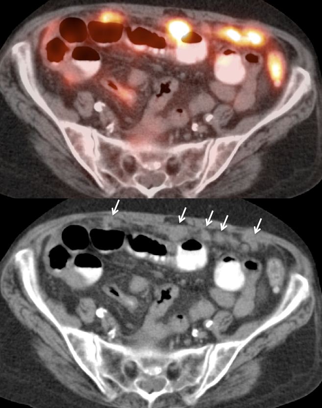
- Peritoneal Carcinomatosis
- Pre-Sacral Soft Tissue After Rectal Surgery
- Gallbladder: Cholecystitis vs. Malignancy
- Inguinal Herniorraphy
- Bladder in Inguinal Canal
- Uterine Fibroid
- Tampon
5. Miscellaneous (continued)
-
Vascular Uptake
- Atherosclerosis
- Vasculitis
- Vascular Grafts
-
Value of NAC PET Images
- Resolving AC Artifacts
- Lung Nodule Identification
-
Non-Malignant, Yet Clinically Significant, FDG-Avid CT Abnormalies
- Acute Diverticulitis
- Colitis
- Cholecystitis
- Pneumonia
- Abscess
- Pancreatitis
- Skeletal Abnormalities [See Chapter 7]
Chapter 7: The Bones
"...is connected to the..."
Chapter 8: The Cancers
"Putting it all together"
Ovarian Cancer
Fast Facts:
- 2nd most common gynecologic malignancy; most lethal.
- 5th leading cause of death in women.
- Advanced disease @ diagnosis (> 50% Stage III)
- 5-year survival: 45 %
Normal Ovarian Uptake:
Premenopausal:
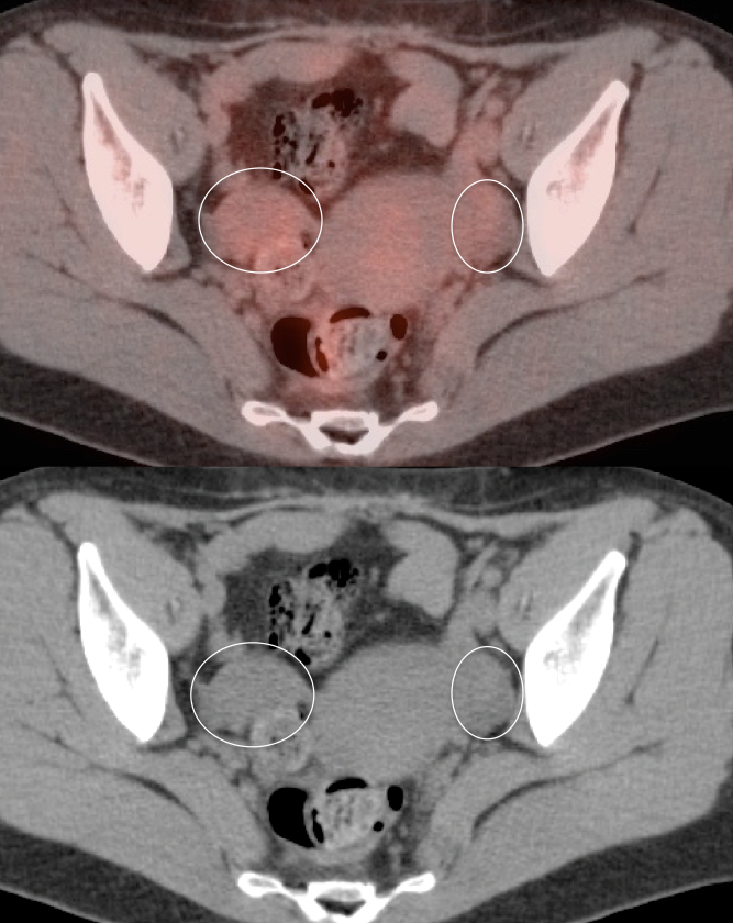
- The normal premenopausal ovary typically demonstrates little or no FDG uptake.
- Moderate and even intense metabolic activity in a premenopausal ovary (unilateral or bilateral) is nearly always a normal finding — in the absence of an associated CT abnormality.
Postmenopausal:
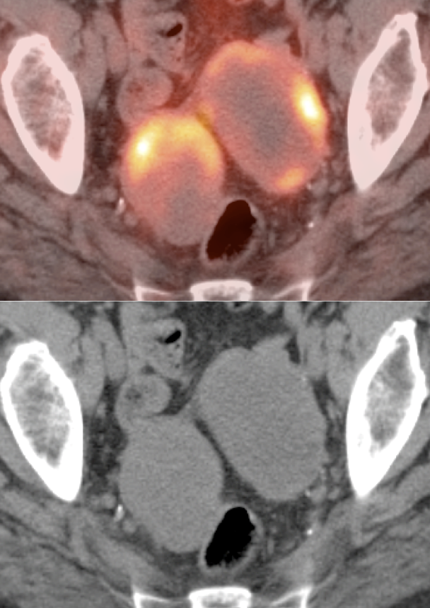
- While the normal postmenopausal ovary can demonstrate varying degrees of metabolic activity, further evaluation is recommended for any postmenopausal ovary with uptake above blood pool background – even in the absence of an associated CT abnormality.
Indications for PET/CT Scan:
Screening:
No current role in the screening of ovarian cancer.
Diagnosis:
- Very limited role in the diagnosis of ovarian cancer, due to significant false positives and false negatives in the evaluation of primary lesions.
- There may be a limited role for PET/CT in the evaluation of equivocal lesions in postmenopausal women.
Initial Staging:
- Initial staging is surgical.
- PET/CT may be of value when neoadjuvant chemotherapy is contemplated.
- The utility of PET/CT for staging lies in its assessment of regional nodes and distant metastatic disease.
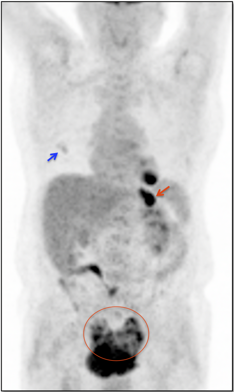
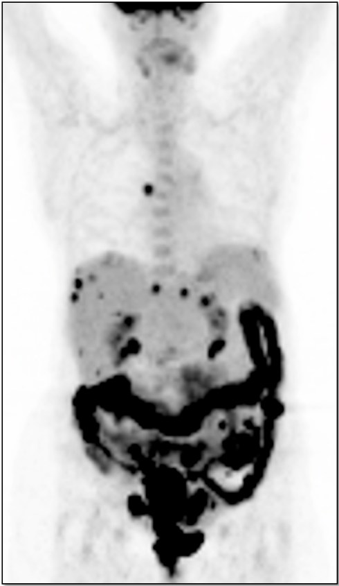
- Distant Disease: Most commonly to the liver, peritoneum, lung, pleura, bones and adrenal glands.
Recurrence & Restaging:
- Restaging suspected recurrence, when CA125 is rising and conventional imaging is negative or equivocal.
- PET/CT alters management in >50% of patients with recurrent disease.
- Distinguishing recurrence from post-therapeutic inflammation.
Assessing Response to Therapy & Prognosis:
- Early Response: Non-responders can be offered alternative therapy (most commonly in the setting of neoadjuvant chemotherapy).
- Late Response: Assess success or failure of therapy, and ultimate outcome/prognosis.
What We Report:
Size, metabolic activity & location of the primary lesion (if present) and representative metastatic lesions.
Primary Lesion:
- Most ovarian cancer patients presenting for PET/CT imaging have already been surgically staged, so the primary lesion will have been resected.
- Pre-surgical scans are occasionally ordered to assess early response to neoadjuvant chemotherapy.
- On occasion, an incidental primary ovarian cancer will be identified.
Metastatic Disease:
- Direct invasion of adjacent structure/organs.
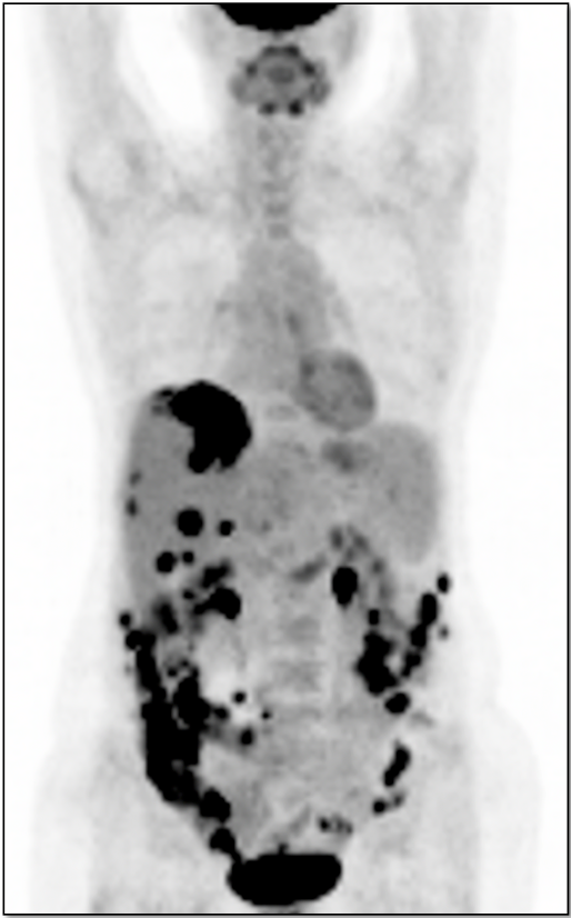
- Seeding of peritoneal cavity.
- Lymphatic spread to pelvic and paraaortic lymph nodes.
- Hematogenous spread, often to liver and lungs.
Ovarian Cysts:
Non-avid ovarian cysts measuring less than 4.0 cm in premenopausal women are presumed functional, as long as solid elements are not present on the co-registered non-contrast CT images (if greater than 4.0 cm, we recommend a 6-week follow up ultrasound, even though they are still likely functional).
We typically report, “A 3.5 x 3.0 cm non-avid left ovarian cyst is noted, presumed functional in a premenopausal woman.“
Ovarian cysts (as are nearly all cysts in the body) should be non-avid. If an ovarian cyst demonstrates FDG uptake (especially is the uptake is focal), ultrasound correlation is recommended.
In a postmenopausal woman, even a non-avid ovarian cyst warrants close attention. We generally report, “A 3.5 x 3.0 cm non-avid left ovarian cyst is present. Although non-avid, the finding of an ovarian cyst in a postmenopausal woman warrants close clinical assessment.”
False Positives:
- Physiologic Uptake
- Inflammatory lesions
- Endometrioma
- Corpus luteum cyst
- Dermoid
False Negatives:
- Lesions < 8.0 mm
- Low-grade, borderline, and some mucinous lesions
- Cystic lesions (only solid elements are FDG avid)
- Low-volume carcinomatosis