
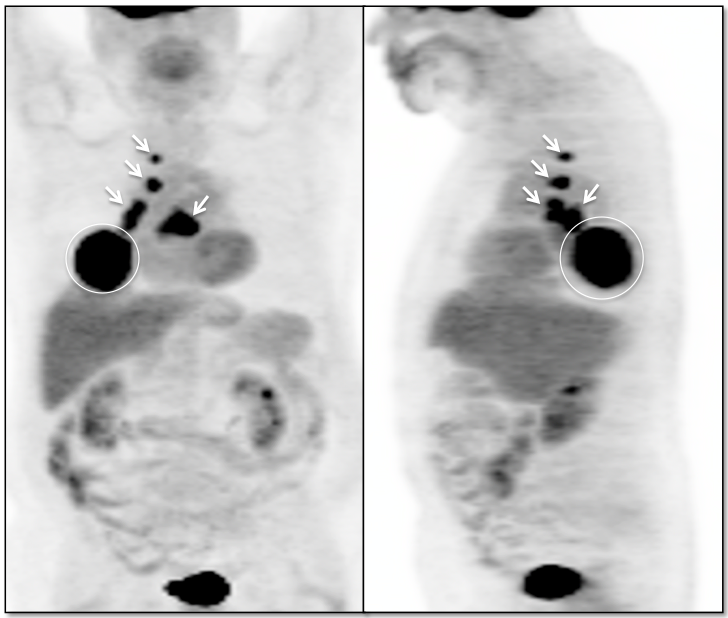
Case 1

Case 1 (Image 2)
Intensely avid right lower lobe pulmonary mass, consistent with a primary lung cancer.

Case 1 (Image 3)
Enlarged FDG-avid metastatic subcarinal lymph node. Intense uptake also involves the right hilum. Although associated enlarged hilar lymph nodes are not visualized on the CT images without intravenous contrast, the appearance is highly suspicious for metastatic right hilar adenopathy.
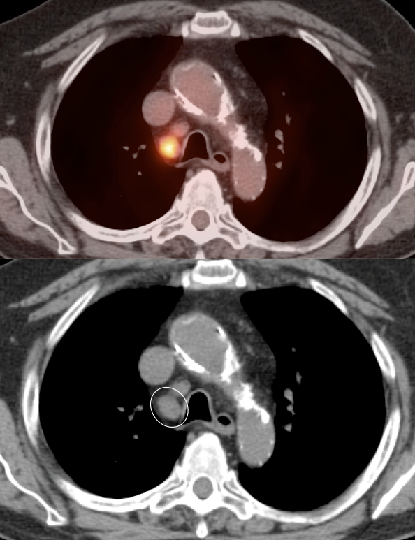
Case 1 (Image 4)
Mildly prominent, intensely avid metastatic right paratracheal lymph node.
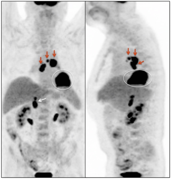
Case 2
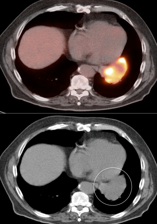
Case 2 (Image 2)
Intensely avid left lower lobe pulmonary mass, consistent with a primary lung cancer.

Case 2 (Image 3)
Intensely avid mediastinal lymph nodes, most consistent with metastatic adenopathy.
Case 2 (Image 4)
Mildly enlarged and intensely avid metastatic subcarinal lymph node.

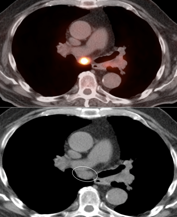
Case 2 (Image 5)
Minimal soft tissue fullness of the right adrenal gland. While its CT appearance is not very impressive, its intense metabolic activity is most consistent with metastatic disease.
Case 3
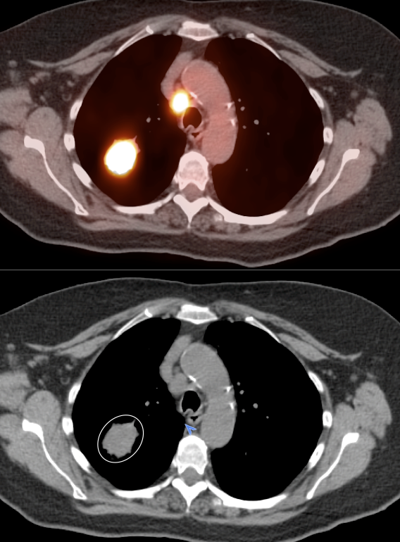
Case 3 (Image 2)
Intensely avid right upper lobe pulmonary mass, consistent with a primary lung cancer.

Case 3 (Image 3)
Enlarged and intensely avid metastatic precarinal lymph node.
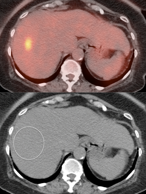
Case 3 (Image 4)
Hypermetabolic liver metastasis. Note that is not apparent on the CT images (a common phenomenon, due to the lack of intravenous contrast administration).

Case 3 (Image 5)
Intensely avid sacral metastasis. An associated osseous abnormality is not clearly present on the CT images. An estimated 50% of FDG-avid osseous metastases are not visible on the CT portion of the exam.

Case 4

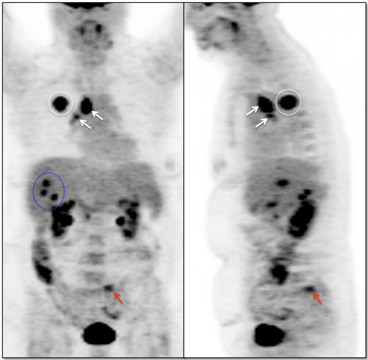
Case 4 (Image 2)
Intensely avid left pulmonary mass, consistent with a primary lung cancer. Adjacent hypermetabolic left hilar and mediastinal nodes are present, consistent with metastatic adenopathy (white arrows).

Case 4 (Image 3)
Enlarged and intensely avid metastatic mediastinal lymph nodes (a portion of the primary lung cancer is visible (circle)).

Case 4 (Image 4)
FDG-avid left adrenal metastasis.

Case 4 (Image 5)
FDG-avid right lung nodule, highly suspicious for a pulmonary metastasis.
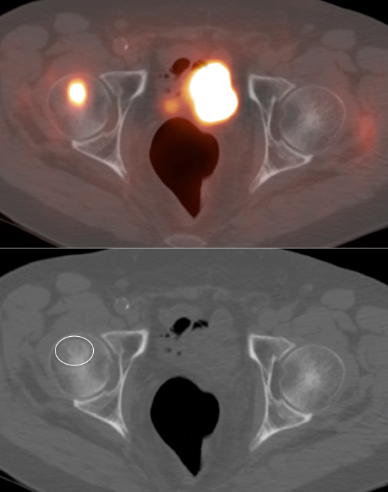
Case 4 (Image 6)
Intensely FDG-avid right femur metastasis (a subtle associated lucency is barely visible on the co-registered CT images).

Case 5
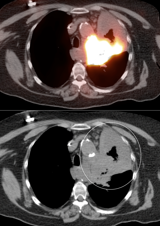
Case 5 (Image 2)
Large region of ill-defined soft tissue density involving the left lung, inseparable from the mediastinum. A large portion of this soft tissue density is intensely FDG-avid, consistent with active malignancy (the non-avid component can represent post-obstructive changes or the sequela of treated disease).

Case 5 (Image 3)
Two hypermetabolic liver metastases.

Case 5 (Image 4)
Several hypermetabolic nodules are noted to involve the subcutaneous soft tissue of this patient, most consistent with metabolically active metastases.
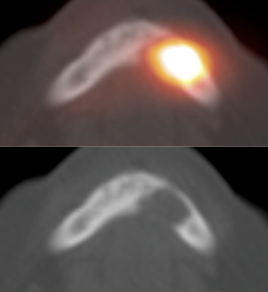
Case 5 (Image 5)
Intensely avid lytic metastasis of the mandible.

Case 5 (Image 6)
Intensely avid lytic metastasis of a vertebral body.

Case 6

Case 6 (Image 2)
Axial images confirm a hypermetabolic primary right lower lobe lung cancer.

Case 6 (Image 3)
Post-treatment scan demonstrates a significant decrease in the size and metabolic activity of this lesion.

Case 6 (Image 4)
The post-treatment MIP demonstrates residual metabolic activity within the patient’s much improved lung cancer. As the residual soft tissue nodule demonstrates metabolic activity equivalent to liver uptake on this follow up study, residual active malignancy is suspected.

Case 7

Case 7 (Image 3)
5-months post chemoradiation. The post-radiation inflammatory changes have improved. The mass is now smaller and demonstrates only minimal uptake.
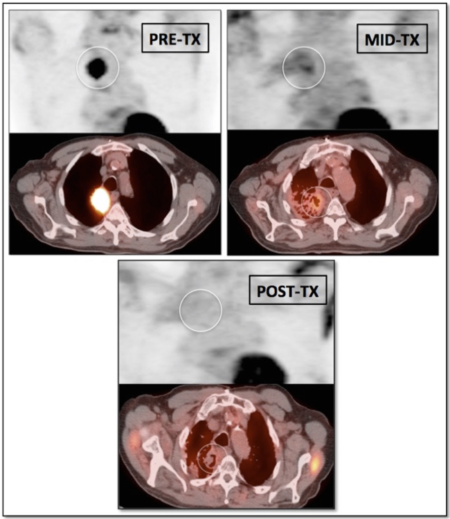
Case 7 (Image 4)
Comparison of the three scans, illustrating the typical effects of therapy on a lung mass and the surrounding pulmonary parenchyma.

Case 8

Case 8 (Image 2)
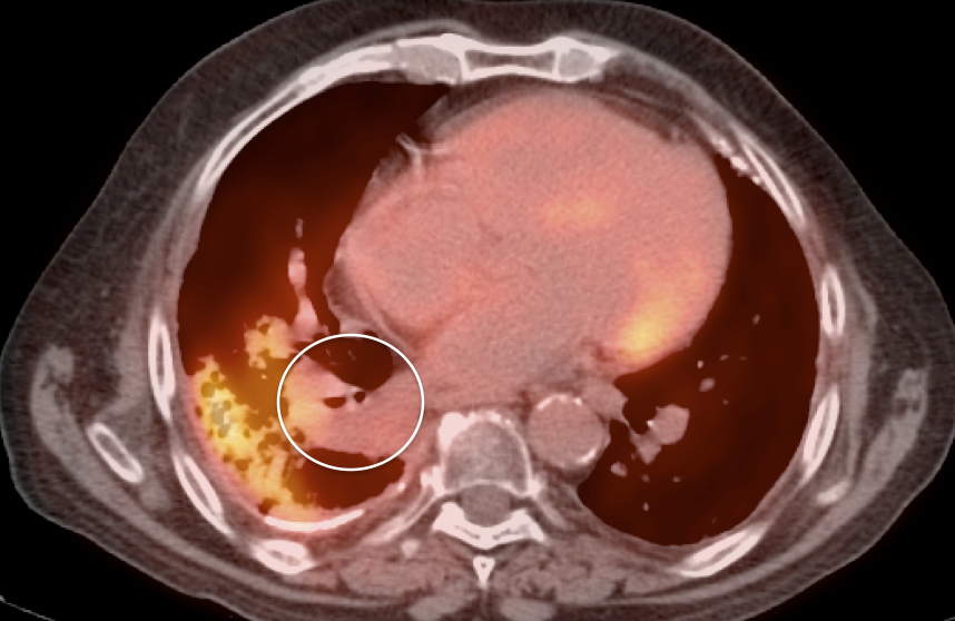
Fused axial image confirms the location of this hypermetabolic primary lung cancer.
Case 8 (Image 3)
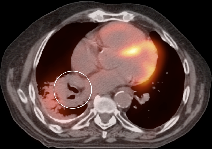
2-months post chemoradiation. The mass is now smaller and more ill-defined. Its metabolic activity has also decreased. Surrounding post-radiation inflammatory changes are present (acute radiation pneumonitis). It is not possible to determine whether the remaining avidity of the mass reflects active malignancy or post-radiation inflammation. Follow up is required.
Case 8 (Image 4)
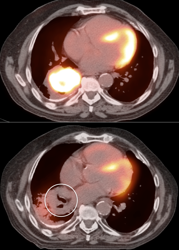
4-months post chemoradiation. Although residual post-radiation inflammatory changes of the pulmonary parenchyma remain, the patient’s primary lung mass is now non-avid, presumed to represent post-therapeutic scarring.
Case 8 (Image 5)
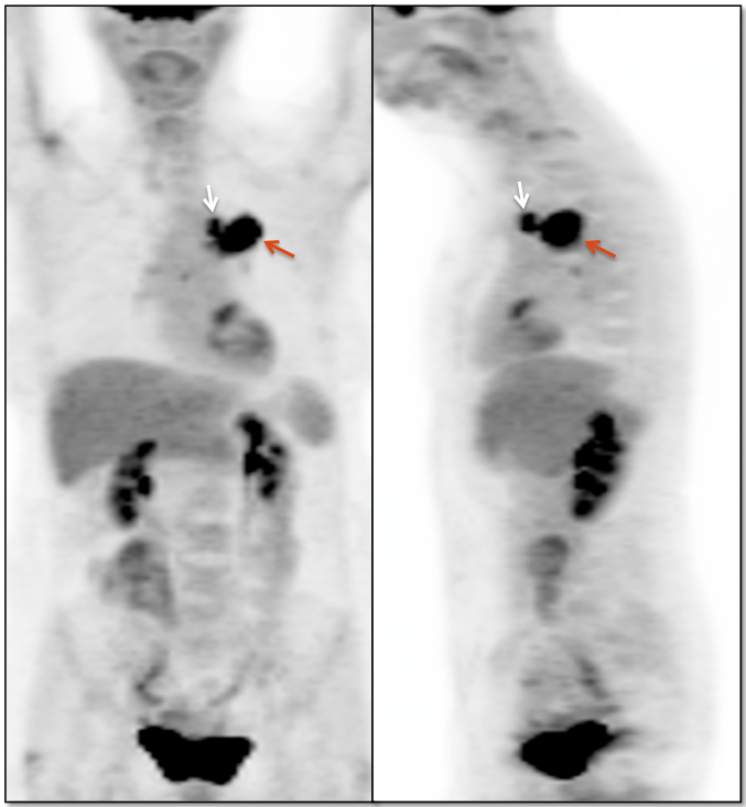
Comparison images of the original hypermetabolic malignancy and the resulting non-avid post-therapeutic scarring.
Case 9
Case 9 (Image 2)
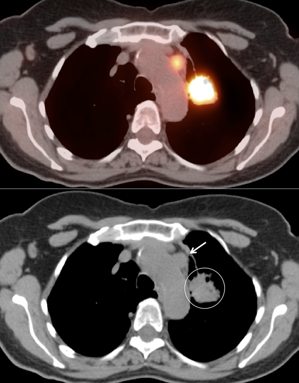
Axial images confirm the hypermetabolic primary lung cancer and an adjacent FDG avid metastatic prevascular lymph node (arrow).

Case 9 (Image 3)
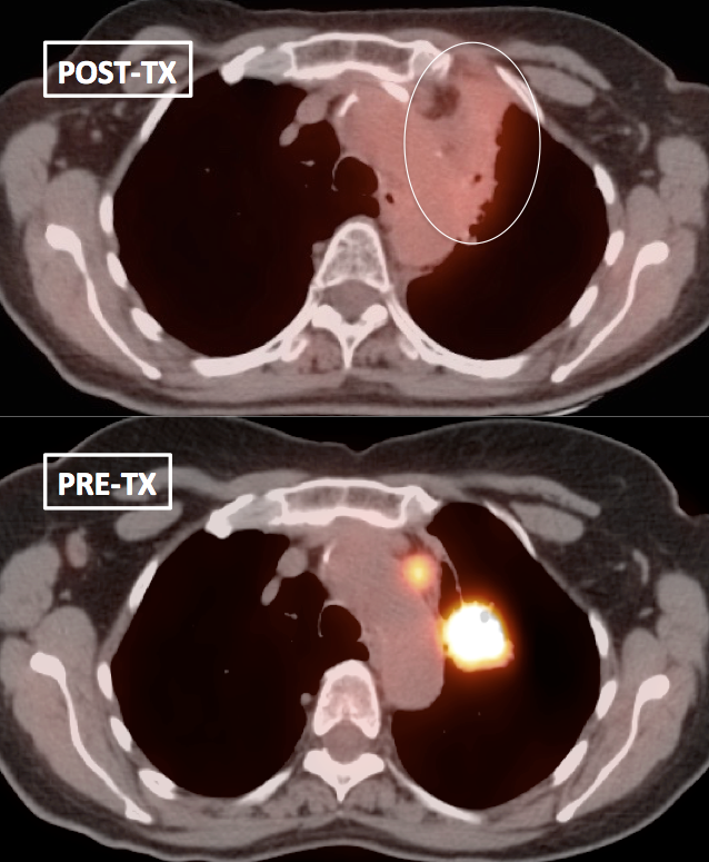
4-months after treatment. The mass and adjacent node have coalesced into prominent, ill-defined soft tissue density. The metabolic activity of these findings has markedly improved. Some residual metabolic activity, however, remains (greater than mediastinal blood pool uptake; equivalent to liver uptake).
Case 9 (Image 4)
Axial comparison of the pre and post therapy scans demonstrates marked improvement, yet residual metabolic activity.

Case 9 (Image 5)
Although comparison MIP images demonstrate marked interval improvement, the residual metabolic activity is still equivalent to liver uptake. So while this may now represent post-therapeutic scarring, residual active malignancy cannot be excluded. Follow up is recommended.

Case 10
Case 10 (Image 2)
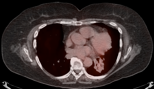
Residual ill-defined soft tissue density in the left lung, not significantly avid, 12-months after radiation treatment of an intensely avid lung cancer, most consistent with post-therapeutic scarring.

Case 10 (Image 3)
Residual ill-defined soft tissue density in the left lung 15-months after radiation therapy of an intensely avid lung cancer, non-avid. The appearance is typical of post-therapeutic scarring.

Case 10 (Image 4)
Residual ill-defined soft tissue density in the left lung 12-months after radiation treatment of an intensely avid lung cancer. This tissue is non-avid and has the typical appearance post-therapeutic scarring.
Case 10 (Image 5)
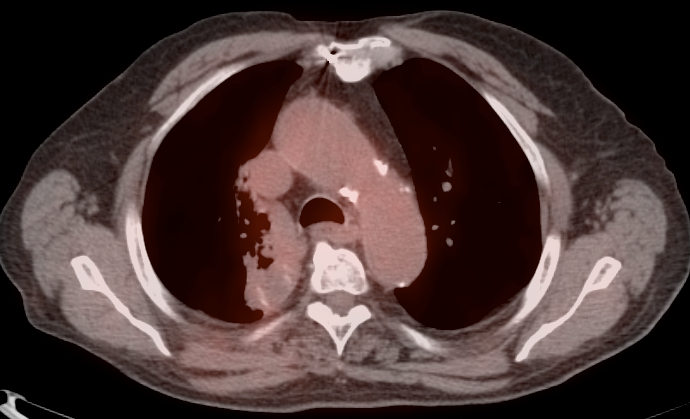
Residual ill-defined soft tissue density after treatment of an intensely avid lung cancer, no longer significantly FDG avid. Note the linear margin of the density, corresponding to the radiation treatment field/port.

Case 11
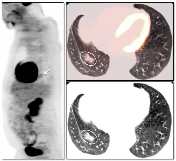
Case 11 (Image 2)
Ill-defined 2.5 cm focal density in the right lower lobe, only mildly FDG-avid. As this density appeared slightly larger than on a prior chest CT scan 3-months previously, biopsy was performed, revealing adenocarcinoma-in-situ (formerly “bronchoalveolar carcinoma”).

Case 11 (Image 3)
1.5 cm nodular density in the left upper lobe lower, a component of which is of ground-glass attenuation. It is only minimally FDG avid, with an SUV of only 0.7 (lean body mass, maximum). The patient desired biopsy, revealing adenocarcinoma-in-situ (formerly “bronchoalveolar carcinoma”).